Our Services • Computed Tomography
Advanced CT Applications
Modern CT captures more than anatomy. With the right scan protocol and specialised post-processing, the scanners we already use can also show how organs are working. Our newest application is CT:VQ™ — ventilation and perfusion mapping of the lungs from a standard non-contrast chest CT.
Now available • August 2026
CT:VQ™ lung ventilation and perfusion studies
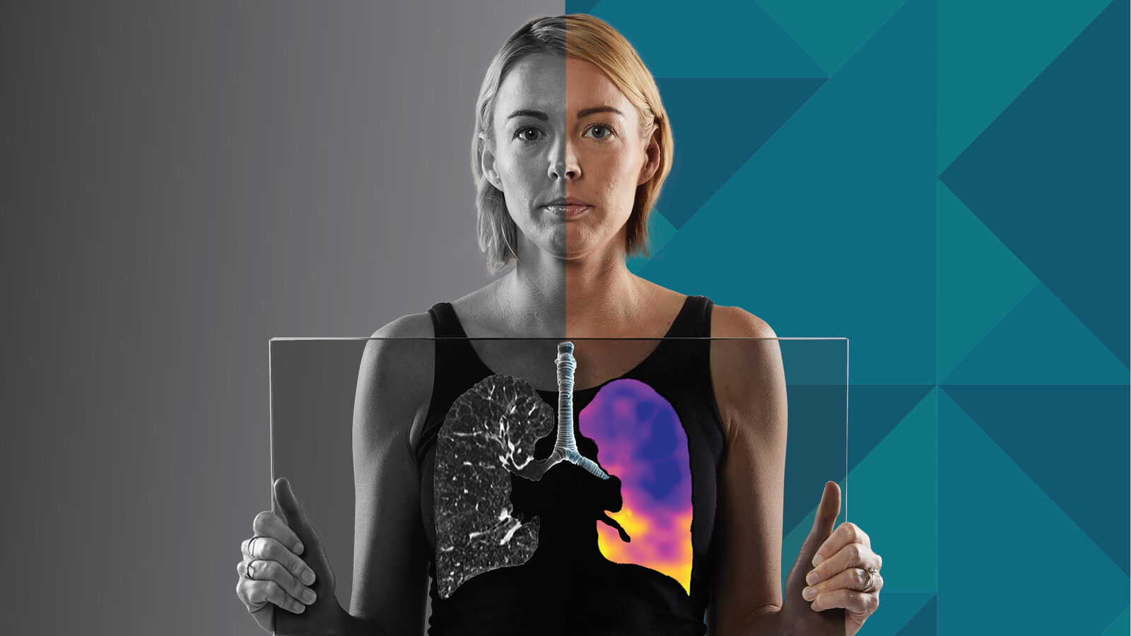
CT:VQ™ is now available at Spectrum Medical Imaging. It is TGA-approved software developed by the Australian company 4DMedical that analyses suitable non-contrast chest CT scans and maps both how air moves through the lungs (ventilation) and how blood flows through them (perfusion).
There are no injections and no radioisotopes involved — it is an extra layer of functional detail drawn from imaging we are already doing. In many cases the analysis can be run on scans that have already been performed, provided the right inspiratory and expiratory series are available.
It is a practical addition to the imaging we already provide, sitting alongside our cardiac CT, CT angiography, low-dose lung cancer screening and other advanced CT services.
How it works
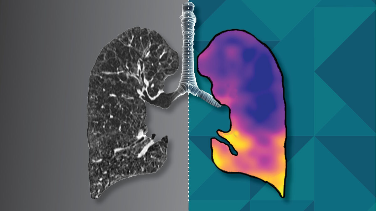
CT:VQ™ is a post-processing step. When we have a suitable pair of inspiratory and expiratory non-contrast chest CT scans — newly performed or already on file — the software generates colour overlays and measurements showing:
- where air is moving (ventilation)
- where blood is flowing (perfusion)
- how the different lobes compare, plus key lung volume measurements
Results come back into our usual reporting workflow and sit alongside the original CT images for the reporting radiologist.
What the report includes
- Colour ventilation and perfusion overlays registered to the CT anatomy, with intuitive scales
- Quantitative metrics, including total lung volumes
- Lobar distribution — size, ventilation and perfusion by lobe, and right-versus-left comparison
- A structured report returned directly into the radiology workflow, alongside the CT report
Who it may help
CT:VQ™ can be useful for patients being assessed before certain lung or thoracic procedures, people with unexplained breathlessness or more complex lung disease, and those who need their lung function monitored over time. It is also an option where traditional nuclear medicine V/Q imaging is not available or not easily accessible.
Not every chest CT is suitable for analysis. Our radiology team determines suitability for each patient, and your referring doctor can discuss whether CT:VQ™ is appropriate for you.
What to expect on the day
If a new scan is needed, CT:VQ™ uses a standard non-contrast chest CT — there is no contrast dye and no injection. Two short scans of the chest are taken during the same appointment:
- You lie on the CT table, usually with your arms above your head if this is comfortable for you.
- The radiographer asks you to take a deep breath in and hold it while the first scan is taken.
- After breathing normally for a moment, you are asked to breathe out as much as you can and hold your breath while the second scan is taken.
The radiographer checks the images after each scan, which may take a few minutes. The appointment usually takes less than 15 minutes. Please wear loose, comfortable clothing without metal buttons, buckles or zips, bring your referral, and tell the radiographer before your scan if you are, or might be, pregnant.
CT uses ionising radiation. The dose is higher than for a standard X-ray, but our scanners adapt the dose to each patient and our radiographers use the lowest dose needed to obtain clear images. Your doctor weighs the benefits of the scan against any potential risks before referring you.
If you have already had a suitable chest CT, there may be nothing further to do — the analysis can often be run on the existing images.
Your results
The CT:VQ™ report is sent to your referring doctor together with the CT report. Please discuss the results with your doctor, who can explain what they mean for you. Patients can view their images through the Spectrum Patient Portal, and referring doctors through our image access service.
For referring doctors
Common applications include CTEPH evaluation, COPD phenotyping, planning for bronchoscopic lung-volume reduction, pre-operative assessment and longitudinal monitoring of lung function.
Full analysis requires paired non-contrast inspiratory and expiratory volumetric chest CT series. Where a patient has a recent chest CT on file, the analysis can often be added retrospectively without a further scan, provided the required series are available — not every historical CT will qualify, and the radiology team will determine suitability.
Please indicate CT:VQ™ — or the clinical question, such as ventilation/perfusion assessment — on your request so our CT team can plan the inspiratory and expiratory series. If you have a question about a particular patient, please contact our CT team.
CT:VQ™ in the news
CT:VQ™ was featured on 7 News in July 2026 following its approval by the Therapeutic Goods Administration.
Frequently asked questions
Do I need an injection for CT:VQ™?
How long does the scan take?
Can CT:VQ™ be run on a chest CT I have already had?
How is CT:VQ™ different from a nuclear medicine V/Q scan?
Will there be any fees involved?

Developed by 4DMedical
CT:VQ™ is developed by 4DMedical, an Australian medical technology company. The analysis is performed on Spectrum’s chest CT images and reported by our radiologists. Learn more at 4dmedical.com
More advanced CT at Spectrum
Cardiac CT
Coronary calcium scoring, CT coronary angiography and TAVI workup CT angiography by our dedicated cardiac radiologists and cardiologists.
Cardiac imagingCT Angiography
CTA of the brain and carotid vessels, aortic CTA for EVAR and TEVAR planning, and embolisation-planning CTA with expert 3D workstation review.
Vascular imagingLow-Dose CT Lung Cancer Screening
Spectrum participates in the National Lung Cancer Screening Program, offering eligible people free, Medicare-funded low-dose CT scans.
About the programCone Beam CT
Dental, oral and maxillofacial imaging with on-site interpretation by our oral and maxillofacial radiologist.
Dental imaging*Please follow preparation instructions as advised by our staff at the time of scheduling your appointment. CT:VQ™ and 4DMedical are trademarks of 4DMedical Limited; CT:VQ™ images on this page are courtesy of 4DMedical. This page is general information and is not a substitute for advice from your doctor.
Ready to book your imaging?
Our team is here to support you and your patients with excellence and care.
Book Appointment